A Rutgers scientist is part of an international team that has determined the process for incorporating selenium—an essential trace mineral found in soil, water and some foods that increases antioxidant effects in the body—includes 25 specialized proteins, a discovery that could help develop new therapies to treat a multitude of diseases from cancer to diabetes.
The research, detailed in Science, includes the most in-depth description yet of the process by which selenium gets to where it needs to be in cells, which is crucial for many aspects of cell and organismal biology. First, selenium is encapsulated within selenocysteine (Sec), an essential amino acid. Then, Sec is incorporated into 25 so-called selenoproteins, all of them key to a host of cellular and metabolic processes.
Understanding the workings of these vital mechanisms in such a detailed manner is critical to the development of new medical therapies, according to researchers including Paul Copeland, a professor in the Department of Biochemistry and Molecular Biology at Rutgers Robert Wood Johnson Medical School.
"This work revealed structures that had never before been seen, some of which are unique in all of biology," said Copeland, an author of the study.
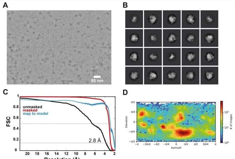
Copeland and the team were able to visualize the cell mechanisms by using a specialized cryo-electron microscope, which uses beams of electrons rather than light to form three-dimensional images of complex biological formations at nearly atomic resolution. The process uses frozen samples of molecular complexes and then applies sophisticated image processing—employing today's vast computing power to string together thousands of images to produce three-dimensional cross-sections and even stop-motion animation conveying a sense of motion within the biomolecules. As a result, scientists can view representations of the intricate structure of proteins and other biomolecules and even how these structures move and change as they function as cellular "machines."
The incorporation of selenium takes place deep within an individual cell's intricate machinery. Scientists already knew which proteins and molecules of RNA—a nucleic acid present in all cells involved in the production of proteins—enabled the process. However, they were not able to discern the critical step of how these factors worked in tandem to complete the cycle, dictating the function of the cell's ribosome—a large macromolecular machine that binds RNA to make more proteins. What they found was that the processes that occur are not like any understood to take place anywhere else in the human body.
"This amino acid gets attached to a unique RNA molecule and that has to be carried to the ribosome via a unique protein factor," said Copeland, whose lab has spent the past 20 years working to understand how these biomolecules function on a biochemical level. "And all of this evolved in humans specifically to allow selenium to be incorporated into this handful of proteins."
Once Sec is ensconced in the selenoproteins, the proteins perform a wide range of vital functions necessary for growth and development. They produce nucleotides, the building blocks of DNA. They break down or store fat for energy. They create cell membranes. They produce the thyroid hormone, which controls the human body's metabolism. And they respond to what is known as oxidative stress by detoxifying chemically reactive byproducts in cells.
Diseases and disorders such as cancer, heart disease, male infertility, diabetes and hypothyroidism can arise when the production of selenoproteins is disrupted.
"Understanding the mechanism by which Sec is incorporated is a foundational part of developing new therapies for a multitude of disease states," Copeland said.
Explore further
Citation: Researchers observe vital cellular machinery behind the body's incorporation of selenium (2022, June 17) retrieved 17 June 2022 from https://ift.tt/v7DKMwS
This document is subject to copyright. Apart from any fair dealing for the purpose of private study or research, no part may be reproduced without the written permission. The content is provided for information purposes only.
"behind" - Google News
June 17, 2022 at 06:14PM
https://ift.tt/v7DKMwS
Researchers observe vital cellular machinery behind the body's incorporation of selenium - Phys.org
"behind" - Google News
https://ift.tt/Tcp9dgz
https://ift.tt/Y270fTn
Bagikan Berita Ini


0 Response to "Researchers observe vital cellular machinery behind the body's incorporation of selenium - Phys.org"
Post a Comment